
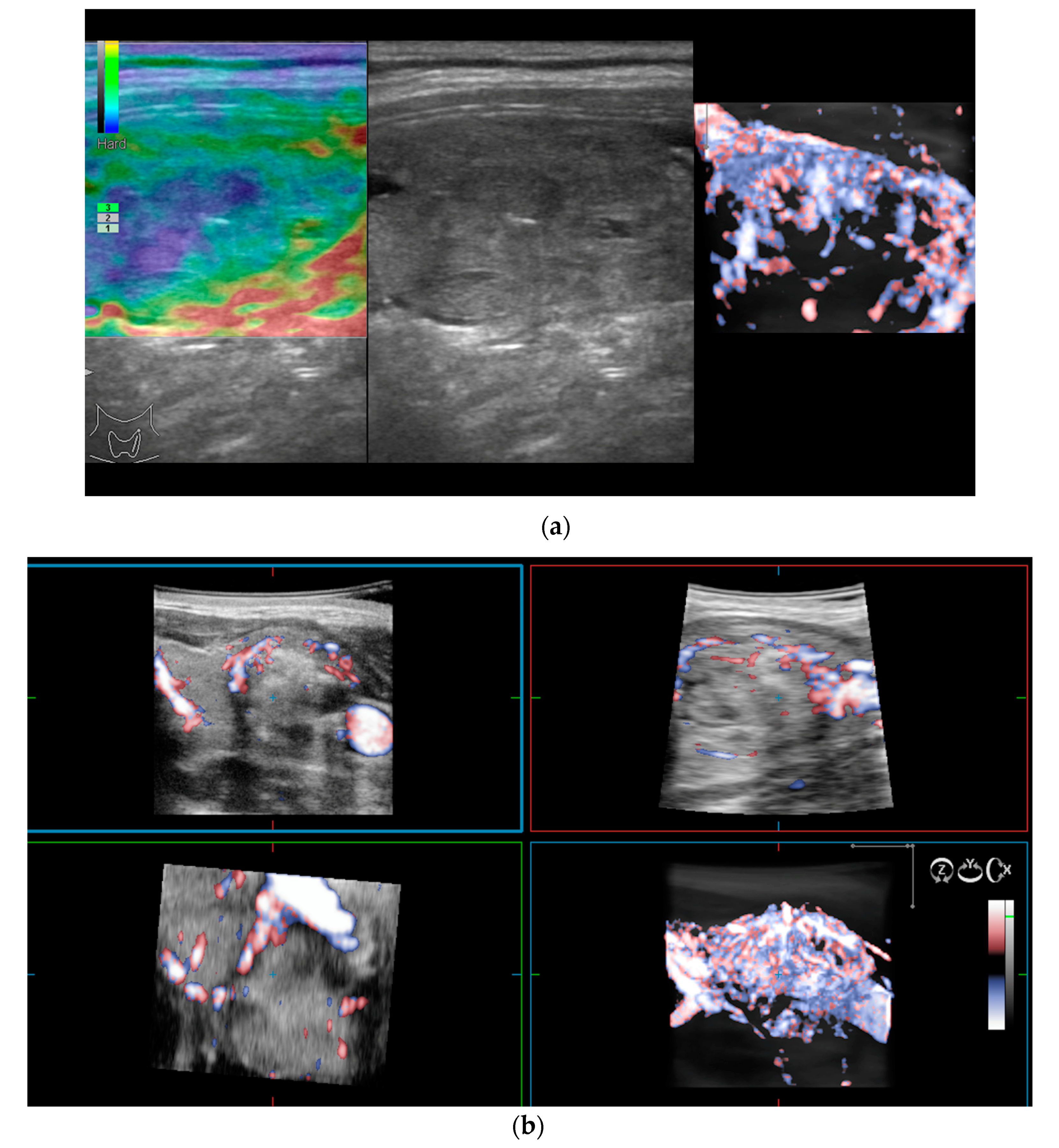
Color flow imaging reveals a spectacular thyroid inferno with marked hyper vascularity. With 3D SMI the vascular structure and vessel branching relationships can be visualized.
Pseudothickening Of The Endometrium In Patient With Adenomyosis A Sagittal Transvaginal Ultrasound Imag Transvaginal Ultrasound Ultrasound Medical Definition
Varicosities stenoses complex interconnecting channels and venous lakes are typical 5 6.

. The term flow void is widely used among radiologists and others involved in MR imaging. There are 3 types of vascularity in thyroid nodules. The use of color flow Doppler CFD or color Doppler imaging CDI or simply color Doppler sonography allows the visualization of flow direction and velocity within a user defined area.
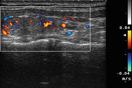
In addition a variance map can tell specific information about flow characteristics - there are four colors. One gradation usually red indicates a flow of blood or tissue in case of color tissue doppler mode moving towards the ultrasound probe and second usually blue indicates a bloodflow or tissue moving from the ultrasound probe but in order to not mistaken you need to look at the legend in the left upper corner of the screen where two colors will be indicated the top one to the ultrasound probe the bottom - from the ultrasound. A region of interest is defined by the sonographer and the Doppler shifts of returning ultrasound waves within are color-coded based on average velocity and direction.
The lumen and back wall are obscured by shadowing from air in the fabric of the covered stent. Further studies are warranted to investigate the predictive role of increased vascularity in diagnosing suspicious thyroid nodules. Is the amount of hue present in a mix with white.
Monochrome SMI mSMI visualizing squamous cell. Many tumors may be classified by using the IOTA Simple Rules model. In contrast to Hashimotos thyroiditis return of normal thyroid appearance is possible at the time of remission.
A careful search for underlying arteriovenous fistulae is necessary. Colour flow imaging shows slow turbulent flow within dilated compressible vascular spaces Fig. Grey-scale ultrasound reveals hypoechoic structures of the vascular spaces.
Color-coded SMI cSMI demonstrates flow and greyscale information with high temporal and spatial resolution simultaneously. Type 2 small amount of internal vascularity in the lesion and a little more concerning. It appears that utilization of vascular flow on color Doppler sonography may not accurately predict malignancy in thyroid nodules.
Color-flow changes are not used to determine specific percent degrees of stenosis other than occlusion. The lesion is hypervascular on color Doppler imaging and demonstrates both arterial and venous waveforms. On MRI T1W imaging demonstrates a fatty mass with intralesional signal voids in keeping with vascular.
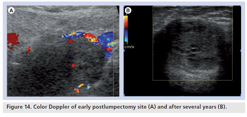
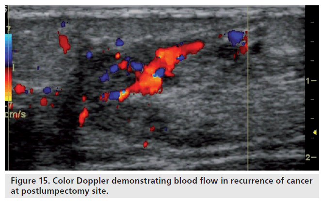
Any breast lesion with radial rather than marginal connecting vessels should be regarded with suspicion 15. Color Doppler evaluation shows diffusely increased vascular flow throughout the testicle. This benign vascular lesion consists of a proliferation of thin-walled vessels lined by flattened endothelial cells within a loosely.
The colors on the left indicate that the flow is laminar. The color score increases with the amount of color flow seen up to a color score of 4 very strong blood flow which can indicate a malignant or M-feature. Type 3 very vascular throughout lesion is the most worrisome and suspicious for malignancy.
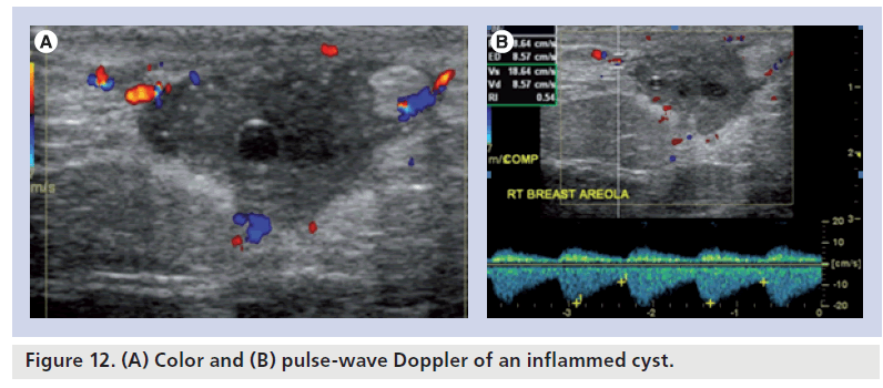
It refers to the low signal seen in vessels that contain vigorously flowing blood and is generally synonymous with vascular patency. Thickening of the cyst walls is seen with gray-scale imaging. 7 Pulsed Doppler shows a marked increase.
CSpectral Doppler waveform reveals very low-velocity flow of 123 cms. Flow voids can also be seen with active flow. Color Doppler ultrasound vascular pattern morphology improves the accuracy and sensitivity of B-mode image diagnosis breast cancers and fibroadenomas with a minimal loss of specificity.
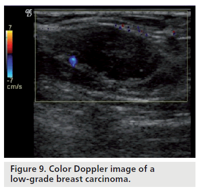
US usually demonstrates a well-defined homogeneously echogenic mass. Also there was no significant difference in internal vascularity between malignant and benign thyroid nodules 95 CI. Echogenic solid mass with marked vascular flow on color Doppler imaging at 8 oclock 10 cm from the nipple measuring 6 x 4 x 6 mm corresponding to the site of the mammographic mass.
Initially this was interpreted as a near TIPS occlusion or high-grade stenosis. Color Doppler demonstrates a ring of vascularity ring of fire pattern around the luteal cyst related to formation of tiny vessels in the walls of the cyst. Type 1 peripheral vascularity or no vascularity and it reflects benign disease.
The use of colour flow Doppler CFD or colour Doppler imaging CDI or simply colour Doppler sonography allows the visualisation of flow direction and velocity within a user defined areaA region of interest is defined by the sonographer and the Doppler shifts of returning ultrasound waves within are colour-coded based on average velocity and direction. However studies on the detection of intra-nodular vascularity in thyroid nodules by color Doppler flow imaging CDFI on intra-nodular vascularity in thyroid nodules have shown conflicting results. The absence of color Doppler flow is assigned a color score of 1 and is considered benign B-feature.
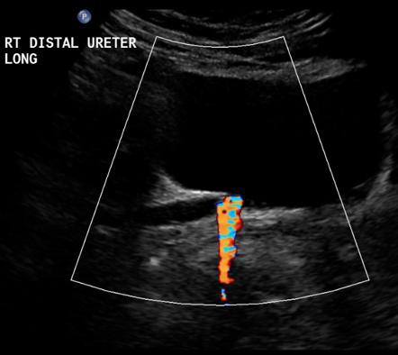
This pattern demonstrates extensive intra-thyroid flow both in systole and diastole. Color flow imaging can be used to detect blood vessels and confirm presence and direction of blood flow whereas spectral Doppler provides more detailed characterization of blood flow and precise velocity measurements to estimate stenosis. Two on top of the map and two on the bottom.
BColor Doppler image demonstrates a thin rim of color in the TIPS lumen just under the near wall. Although most hepatocellular carcinomas have internal vascularity on color flow Doppler images a significant number of metastases also have internal vascularity. Color flow is particularly useful for evaluating the iliac arteries the popliteal trifurcation and the tibial vessels.
The presence of colour Doppler flow was demonstrated in 836 of normal lymph nodes compared with 875 of metastatic lymph nodes. The patient underwent left orchiectomy and the pathology revealed a mixed germ cell tumor with 80 seminoma and 20 embryonal carcinoma component. Color on ultrasound most often relates to blood flow or more specifically movement.
Also color flow can identify the presence and length of an arterial occlusion as well as the distal site of reconstitution. One color will represent flow in the veins while the other color will represent flow in the arteries depending on the direction of flow in relation to the probe its a physics thing. This overlap limits the usefulness of color flow Doppler imaging for distinguishing hepatocellular carcinoma.

Pin On Radiology Signs

Pin On Thyroide
Color Doppler Sonography Characterizing Breast Lesions

Pin Van Dr Abuaiad Op Brain Head And Neck
Doppler Ultrasound Radiology Key
Pin On Thyroide
Color Flow Doppler Ultrasound Radiology Reference Article Radiopaedia Org
Diagnostics Free Full Text Ti Rads Diagnostic Performance Which Algorithm Is Superior And How Elastography And 4d Vascularity Improve The Malignancy Risk Assessment Html

Dx Osseous Metaplasia Of Endometrium Abstract Artwork Abstract Artwork

Color Doppler Patterns A Pattern 0 Normal Thyroid Vascularity B Download Scientific Diagram
Color Doppler Sonography Characterizing Breast Lesions

Color Doppler Imaging Of The Appendix Xu 2016 Journal Of Ultrasound In Medicine Wiley Online Library

Arteriovenous Malformation Of The Thigh A Color Doppler Showing A Download Scientific Diagram

Pin On Thyroid

Epos Trade
Color Doppler Sonography Characterizing Breast Lesions

Pin On Thyroide
Color Doppler Sonography Characterizing Breast Lesions
Color Doppler Sonography Characterizing Breast Lesions
